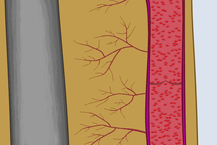
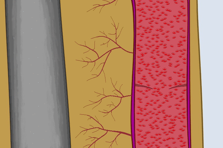
Veins become varicosed when they get widened and the valves are then unable to close effectively. When they close effectively, they are able to hold and push blood forward. When they cannot close effectively, the blood flows backward as illustration here
Spider Veins: These are tiny thin veins that are on the skin’s surface. They may be like red, blue, or green webs just beneath the skin. They are nonfunctional and considered “dead-end” veins. While they don’t usually cause any discomfort, the deeper veins that often accompany them do. These veins are treated using Sclerotherapy which is done by microinjecting them with Polidocanol which shuts them off and the body absorbs them over time. Results are usually noted in about 4 to 6 weeks. Treatment for these veins are considered cosmetic and are not covered by insurance.
Varicose veins are enlarged veins that often appear ropey and may look blue or red. They may also occur deeper under skin and may not be visualized, but are seen by ultrasound. Due to dilation of the vessel, the valves are unable to function which then causes backward flow and accumulation of blood in the lower parts of the legs due to gravity. Most symptoms therefore, associated with varicose veins, are at their worst at the end of the day after being on ones feet.
As the veins worsen, signs and symptoms progress. Usual signs and symptoms associated with varicose veins are pictorially illustrated here.
These veins, depending on size, are treated using either Ultrasound Guided Sclerotherapy, heat inducing Radio Frequency (RF), heat inducing LASER, Mechanical-Chemical ablation, or Microphlebectomy. All these methods have significantly high success rates and are all performed as in office procedures under sterile conditions. There is no down time from these procedures and we require you to return to your normal activities after you leave our office.
Ultrasound Guided Sclerotherapy (USG) consists of using ultrasound for guidance to inject non trunkal veins usually smaller than 7 mm. The vessel is shut almost instantly and is viewable live on screen.
Thermal methods include the heat inducing Radio Frequency (RF) or LASER. A catheter is introduced into a trunkal vein using local anesthesia and then heat is applied which shuts down the vessel. Blood flow is redirected to other veins.
Mechanical-Chemical ablation (MOCA) utilizes a small catheter that is inserted into a trunkal veins using anesthesia and then turned on to rotate rapidly inside the vein which destroys the inner lining of the vessel. Polidocanol is injected simultaneously which further contributes to the destruction.
Microphlebectomy is a office procedure where veins are stripped out using micro puncture sites. This along with the old fashioned in hospital stripping remain effective methods when needed.